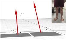
Finite element analysis has gained popularity in anthropological research to connect morphological form to measurable function but requires that loads are applied at appropriate anatomical locations. This is challenging for the ankle because the joint surfaces are not easily determined given their deep anatomical location. While the location of the talonavicular and subtalar joints can be directly determined via medical imaging, regression equations are a common, less invasive method to estimate joint locations from surface anatomy. We propose a regression-based method to locate the in vivo positions of the talonavicular and subtalar joints employing three-dimensional (3D) surface markers, such as those used routinely in gait studies.
Navicular height was measured on weight-bearing radiographs (WBR) and simulated weight-bearing computed tomography (SWCT) scans to ensure SWCT correctly simulated foot weight-bearing configuration. The location of external foot markers and internal locations of the talonavicular and posterior subtalar joint were measured on each SWCT. Stepwise regression analysis was used to select the external markers that best predicted the three internal locations.
Navicular heights measured on WBR and SWCT scans were not statistically different (p = .44), indicating that SWCTs recreate the weight-bearing position of the foot. The navicular tubercle and medial and lateral malleoli were the best predictors of subtalar and talonavicular joint locations. These palpable anatomical locations explained more variation in internal joint location (r2 > .79; SEE < 3.0 mm) than other landmarks.
This study demonstrates that external palpable landmarks can predict the location of the talonavicular and subtalar joints.
